esophagus anatomy skeletal muscle
“Tremendous Tube – GI System, Part 2” – Outlander Anatomy. 9 Images about “Tremendous Tube – GI System, Part 2” – Outlander Anatomy : Esophagus: Anatomy, sphincters, arteries, veins, nerves | Kenhub, Pin em Histology and also “Tremendous Tube – GI System, Part 2” – Outlander Anatomy.
“Tremendous Tube – GI System, Part 2” – Outlander Anatomy
 www.outlanderanatomy.com
skeletal pdhpe tremendous tissues cram memrise describe
Pin Em Histology
 www.pinterest.co.kr
squamous stratified epithelium keratinized non esophagus histology muscle skeletal smooth tissue slides anatomy human cell cells body basal last 1st
Ligament Of Treitz: Suspensory Ligament Of Duodenum | Kenhub
quadratus lumborum diaphragm phrenicus ligament nervus treitz diaphragma crus psoas kenhub zwerchfell vena musculus abdomen cava becken ligaments inferior dextrum
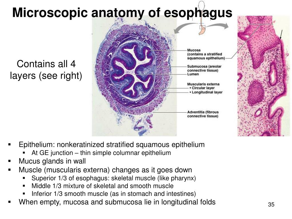
PPT - The Digestive Tract PowerPoint Presentation, Free Download - ID
 www.slideserve.com
esophagus digestive tract microscopic histology layers ppt powerpoint presentation anatomy
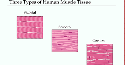
Human Anatomy And Physiology: Skeletal, Cardiac, And Smooth Muscle
 humanphysiologyanatomy.blogspot.com
cardiac skeletal smooth muscle
Jejunum
 www.eugraph.com
histology jejunum intestine epithelia
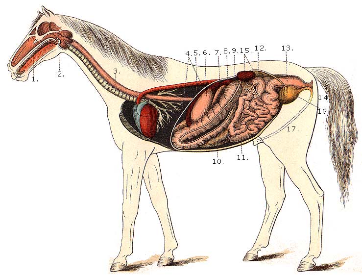
Horse Anatomy Pictures-Think Like A Horse-Rick Gore Horsemanship
 thinklikeahorse.org
horse digestive system anatomy diagram kidney horses mouth equine psoas diaphragm liver stomach spleen bladder muscles oesophagus intestine colon urethra
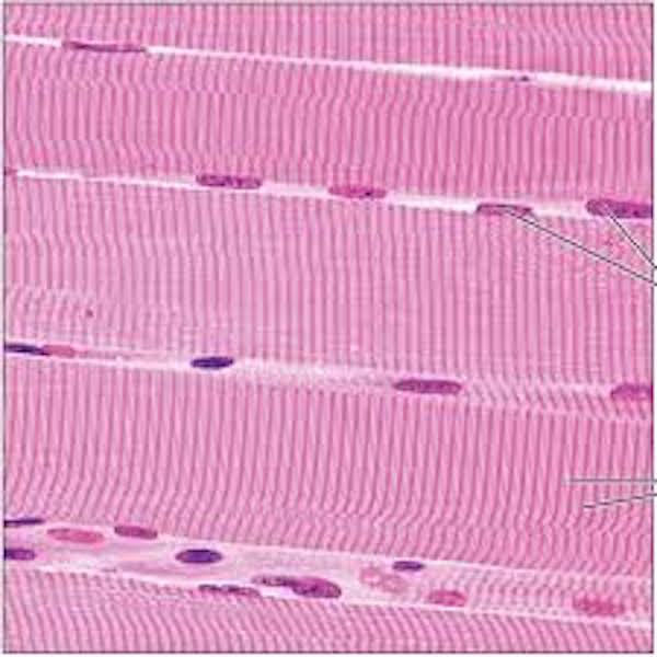
STRIATED, SKELETAL MUSCLE | Microanatomy Web Atlas | Gwen V. Childs, Ph.D.
 microanatomy.net
skeletal striated microanatomy striations
Esophagus: Anatomy, Sphincters, Arteries, Veins, Nerves | Kenhub
esophagus aorta branches abdominal anatomy thoracic arch aortic anterior kenhub situ structures thorax arteries nerves cavity overview veins ventral neurovasculature
Ligament of treitz: suspensory ligament of duodenum. Horse digestive system anatomy diagram kidney horses mouth equine psoas diaphragm liver stomach spleen bladder muscles oesophagus intestine colon urethra. Squamous stratified epithelium keratinized non esophagus histology muscle skeletal smooth tissue slides anatomy human cell cells body basal last 1st